Fetal liver hematopoietic stem cell niches associate with portal vessels
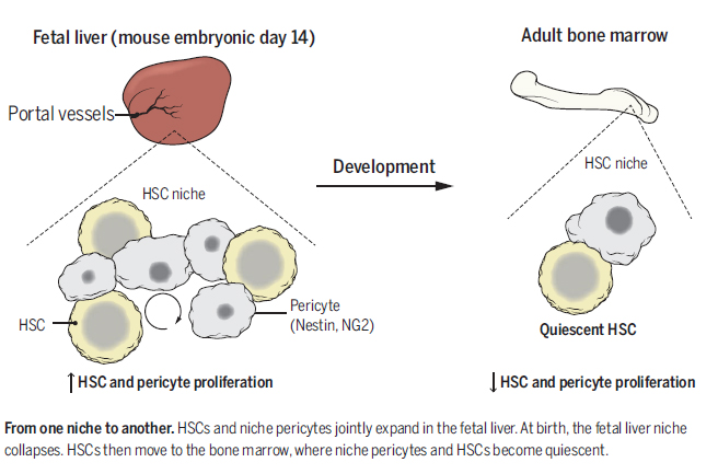
Whereas the cellular basis of the hematopoietic stem cell (HSC) niche in the bone marrow has been characterized, the nature of the fetal liver niche is not yet elucidated. We show that Nestin+NG2+ pericytes associate with portal vessels, forming a niche promoting HSC expansion. Nestin+NG2+ cells and HSCs scale during development with the fractal branching patterns of portal vessels, tributaries of the umbilical vein. After closure of the umbilical inlet at birth, portal vessels undergo a transition from Neuropilin-1+Ephrin-B2+ artery to EphB4+ vein phenotype, associated with a loss of periportal Nestin+NG2+ cells and emigration of HSCs away from portal vessels.These data support a model in which HSCs are titrated against a periportal vascular niche with a fractal-like organization enabled by placental circulation.
Hematopoietic stem cells (HSCs) are generated in the mouse fetus around embryonic day 10.5 (E10.5) from hemogenic endothelium of the dorsal aorta (1, 2), then migrate to the placenta via the umbilical arteries (3) and return to the fetus via the umbilical vein (4). The umbilical vein delivers oxygenated blood to the fetus via the portal sinus, whose branches give rise to the portal vessels in the fetal liver (FL).
In this organ,HSCs undergomarked expansion (5). The predictable growth curve of HSCs during development
suggests that hitherto unknown determinants set the numbers of these cells. See More : www.sciencemag.org
Source : Science Magazine - 8 January 2016


